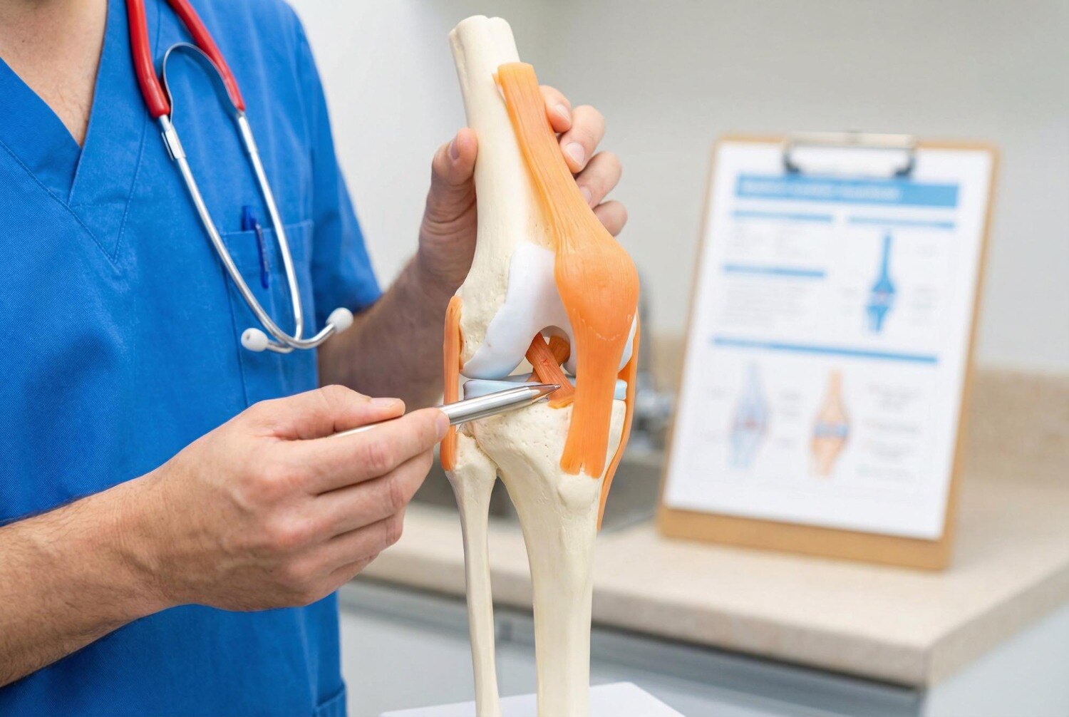
A knee that gives way on stairs, while pivoting, during a run or when rising from a chair needs more than a location-based search for surgery. You will learn how clinicians confirm true instability, which ligament may be involved, when rehabilitation is reasonable, and when reconstruction becomes a practical option.
Key takeaways
- Urgent care is needed after severe pain, deformity, numbness or a cold, pale foot.
- MRI and physical tests help identify the injured ligament and associated cartilage damage.
- Rehabilitation may be enough for some injuries; repeated buckling can support reconstruction.
- Compare graft type, surgeon experience, rehabilitation access and follow-up before choosing care.
What knee instability feels like—and when it needs urgent attention
True knee instability feels like the joint suddenly buckles, shifts or gives way under your weight. You may notice it on stairs, while turning, running, or rising from a chair; fear of another buckle can make you avoid normal movement.
One painful twist does not prove a ligament tear. Arrange an examination if giving-way returns, swelling appears quickly after an injury, the knee cannot fully straighten, it locks, or you cannot walk normally after 24 to 48 hours. Repeated instability can damage the meniscus and cartilage, even when pain settles.
Seek emergency care after a major injury if the knee looks deformed, you cannot bear weight, or the foot becomes cold, pale, numb or weak. Get urgent medical review for these warning signs:
- Severe swelling that develops rapidly, or pain that keeps escalating
- Fever with a red, hot knee or fluid draining from a surgical wound
- New calf swelling or pain
- Chest pain or sudden breathlessness
A clinician should identify the injured structure before anyone discusses ligament reconstruction near ITI Layout. The injury history, stability tests, range of motion and imaging determine whether you need observation, rehabilitation, urgent treatment or a surgical opinion. Search terms such as knee ligament treatment near ITI Layout describe a location, not a diagnosis.
How doctors identify the injured knee ligament
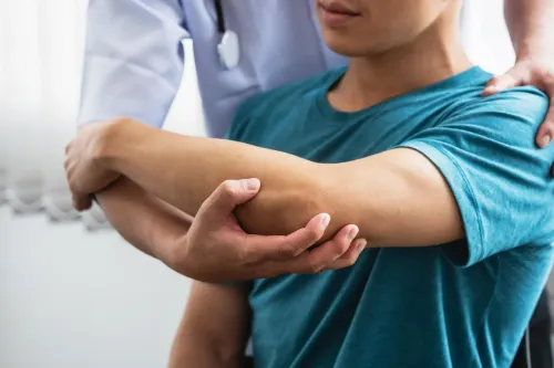
The injured structure is identified by the injury mechanism, swelling pattern, range of motion and targeted stability tests—not by giving way alone. Lachman and pivot-shift tests assess forward movement and rotational instability from an ACL injury; the posterior-drawer test checks backward movement from a PCL injury.
| Structure | Examination findings | Imaging clues |
|---|---|---|
| ACL | Lachman feels soft or excessively loose; pivot shift reproduces a slipping sensation | MRI shows ACL fibre disruption and associated meniscus or bone bruising |
| PCL | Posterior drawer shows the tibia sagging backward; pain followed a dashboard impact or fall onto a bent knee | MRI shows PCL damage and possible cartilage or meniscus injury |
| MCL | Valgus stress causes pain or opening on the inner side of the knee | MRI grades the inner ligament tear and detects nearby injury |
| LCL | Varus stress causes pain or opening on the outer side | MRI assesses the LCL and posterolateral corner |
| Combined | Marked looseness in more than one direction, severe swelling or dislocation history | MRI maps multiple tears; urgent assessment checks blood flow and nerve function |
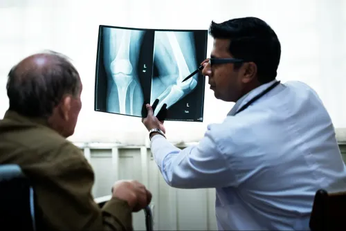
A plain X-ray remains important because it can reveal fracture, dislocation, arthritis or a small bone avulsion that MRI does not replace. MRI then defines the ligament tear and associated damage.
Ask the clinician to document each injured structure before choosing knee ligament treatment near ITI Layout; a treatment plan for an isolated ACL tear differs from one for a combined injury.
When rehabilitation is enough and when reconstruction makes sense
Compare structured rehabilitation with reconstruction when instability persists, your activities demand pivoting, or a meniscal injury changes the stakes—not simply because an MRI shows a tear.
| Option | What it involves | When it fits |
|---|---|---|
| Rehabilitation and activity changes | Progressive strength, balance and neuromuscular training; avoid cutting, jumping or uneven-ground tasks during recovery | Lower-demand lifestyle, an isolated ACL tear without giving-way episodes, or stable isolated MCL injury |
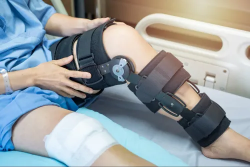
| Brace plus rehabilitation | A hinged brace protects healing tissue while exercises restore movement and quadriceps control | Selected MCL or LCL injuries, or temporary protection after an acute injury; a brace does not replace rehabilitation |
| Reconstruction | Graft surgery followed by staged rehabilitation lasting months | Recurrent giving way, pivoting sport or work, symptomatic functional instability, or an associated meniscal injury |
For an acute isolated ACL tear, early reconstruction is not automatically superior. Initial rehabilitation with the option of delayed surgery produced similar patient-reported outcomes at two years in a randomized trial, although some people later needed reconstruction. PCL, LCL or combined injuries require a structure-specific plan; multiple-ligament instability needs prompt specialist assessment.
Before choosing ligament surgery in Bangalore, ask the clinician to document the injured ligament and associated damage, explain graft choices, name the physiotherapy provider, and set out follow-up and complication plans. “Ligament reconstruction near ITI Layout” is a location search, not a diagnosis. Choose treatment after the examination, not the postcode.
Repair, reconstruction and graft choice are different operations
Repair works when the torn ligament ends remain attached, healthy enough to heal, and reachable for secure stitching. Reconstruction replaces an irreparable or unreliable ligament with a tendon graft; it is not simply sewing the native tissue back together.
| Operation | What happens | When it fits |
|---|---|---|
| Repair | The native ligament is reattached or stitched | A selected tear with viable tissue, including some proximal ACL or collateral-ligament injuries |
| Reconstruction | A graft is fixed to restore the ligament’s path and tension | A stretched, shredded or retracted ACL that will not stabilise the knee reliably |
An ACL is more often reconstructed because it lies inside the knee, where complete tears can retract and lose useful structure. Ongoing giving-way can then injure the meniscus and cartilage. Common autograft sources include:
- Patellar tendon: strong fixation, but kneeling pain can occur
- Hamstring tendon: less kneeling pain, with different strength considerations
- Quadriceps tendon: useful graft size, with its own front-of-knee recovery demands
Autograft is frequently favoured for young or active patients; an allograft from donated tissue suits selected situations and is not automatically equivalent.
Do not choose surgery from the phrase ligament reconstruction near ITI Layout or from “ACL” alone. Ask the clinician to document the injured structure, associated damage, graft options, and why repair or reconstruction fits your knee. The same checks apply when comparing knee ligament treatment near ITI Layout.
What recovery involves and how to compare local surgical care
Do not choose on location alone. A provider for knee ligament treatment near ITI Layout should name the injured structure—ACL, PCL, MCL, LCL or multiple ligaments—and review the MRI with you. Ask whether meniscus tears, cartilage damage, fracture or dislocation changes the operation or its urgency.
Check these points before booking ligament surgery in Bangalore:
| Check before choosing | Ask for | Warning sign |
|---|---|---|
| Diagnosis and graft | The named ligament, graft type, fixation method and reason for that choice | “Standard surgery” without your scan findings |
| Rehabilitation | A staged plan covering motion, swelling, strength, running and return-to-sport testing, plus the physiotherapist’s name | No written milestones or no clear rehabilitation provider |
| Associated injuries | An explanation of meniscus or cartilage treatment during, before or after reconstruction | MRI findings are not discussed |
| Follow-up and safety | Appointment timing, wound review, motion checks and a contact plan for complications | No named clinician for postoperative concerns |
Recovery is measured in months, not weeks. Confirm who checks quadriceps weakness, stiffness and graft progress, and what happens if motion does not return or instability persists.
Discuss infection, blood clots, nerve or blood-vessel injury, graft failure and loss of motion in plain language. Fever, wound drainage, calf swelling, chest pain or sudden breathlessness needs urgent medical review.
At Leela Health Care Orthopaedic Clinic Banashankari, use the consultation to verify these details rather than assuming a multi-specialty clinic automatically handles complex knee reconstruction.
Related service
ORTHOPEDIC Ligament Reconstruction in Banashankari, Bangalore Dr Prajwal KN, Orthopaedic Sports Injury Specialist If you need ligament reconstruction in... View service → |
Frequently asked questions
What does knee instability feel like?
The knee may suddenly buckle, shift or give way during stairs, turning, running or standing from a chair.
How do doctors identify an injured knee ligament?
Doctors combine your injury history with knee stability tests and imaging such as X-rays or an MRI.
When is rehabilitation enough for knee instability?
Rehabilitation can suit selected injuries when the knee remains stable and you can regain strength, movement and function without repeated buckling.
When does ligament reconstruction make sense?
Reconstruction becomes a consideration when instability continues, the knee repeatedly gives way, or you need reliable stability for work or sport.
What is the difference between ligament repair and reconstruction?
Repair reconnects or preserves the damaged ligament when its tissue and attachment allow it; reconstruction replaces it with a graft.
Keywords